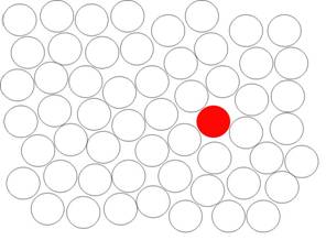
The Brown Location Test was initially designed specifically to measure visual memory difficulties associated with right temporal lobe epilepsy or the impact of removal or damage to the right medial temporal lobe.
The BLT forms A and B have clinical norms for ages 17 through 89. It is available for purchase for clinical or research use at a reasonable cost. It may be available for significantly reduced cost for research purposes. There are hand administered or computer administered versions.

There are no drawing demands and rather brief and simple instructions. The hand administered version is presented in an easy to use flip book format. The directions for use are integrated into the easy to use answer sheet. The use of simple locations rather than objects further reduces verbal mediation to make it a pure visual memory test. The marker chips are large and easy to hold for people with movement difficulties. The computer version only requires the use of a mouse to point and click at the correct locations.
Click here to order the Brown Location Test. Click here to learn more about some of the publications and presentations that have used it.
It has five learning trials in which the same 12 red dot locations are presented for each trial. This is followed by an interference trial with black dots and a short delay free recall trial for the original 12 red dot locations. After a 20 minute delay, the person is again asked to indicate the red dot locations with marker chips. This is followed by a rotated free recall, and then a recognition format.
It demonstrated a clear nonvisual memory factor in factor analysis, has strong psychometric properties, and was found to be strongly associated with damage/removal of the right hippocampus.
In addition, BLT test performance is currently being examined in patients with cancer, Multiple Sclerosis, Traumatic Brain Injury, ADHD, Stroke, OCD, AD/HD, and Autism Spectrum Disorder.
Though designed to test visual memory, case studies and preliminary data suggest that the rotated delay trial seems to be particularly sensitive to the effects of attention difficulties and is typically lower with those who have experienced TBI or have a history of AD/HD.
